Published in : Oncology | January 7, 2026 |
![]() Medically Reviewed
Medically Reviewed
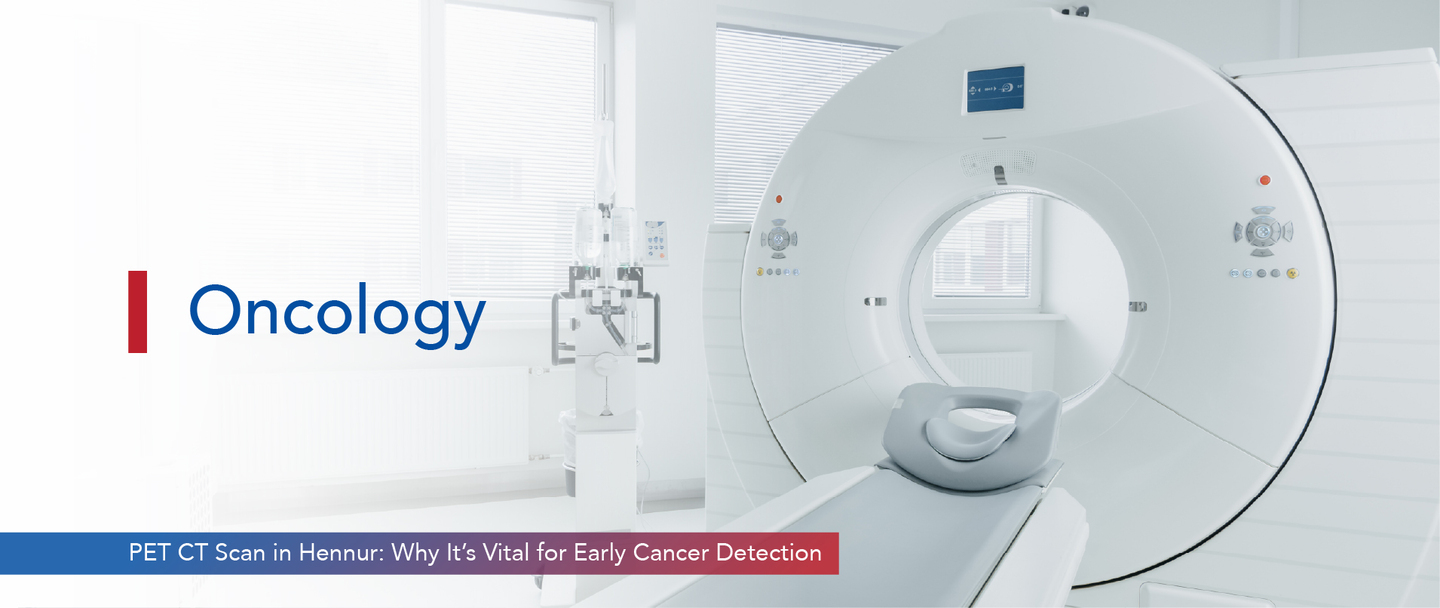
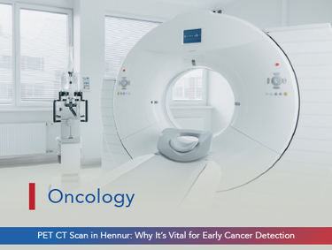
Detecting cancer at an early stage may significantly improve the success of treatment! The sooner a disease is recognised, the more options remain open. One of the most reliable technologies for this purpose is the PET CT scan, which combines two powerful imaging techniques into a single diagnostic tool. When cancer symptoms are not yet clear, and conventional imaging results offer limited insights, the PET CT scan for cancer becomes essential.
People looking for advanced cancer diagnosis tests are now considering locations that offer specialised services. For individuals residing in or near Hennur, timely access to an oncology imaging centre in Hennur may play a crucial role in initiating the right course of action. Early cancer detection remains a cornerstone in cancer management, and the PET CT scan provides just the right level of detail at the right time.
A PET CT scan is a diagnostic test that merges functional and structural imaging. It allows doctors to visualise not just the physical structure of organs but also the metabolic processes occurring within them.
Medical professionals often recommend this scan when high precision is required. A PET CT scan in Hennur offers this combined advantage through one unified diagnostic session.
The PET CT scan is performed when identifying structural abnormalities alone may not be sufficient. It is designed to detect cellular activity that precedes any visible damage in tissues or organs.
Those undergoing complex cancer diagnosis tests often benefit from this kind of detailed imaging. The presence of a PET CT scan Hennur facility makes such testing accessible within the region.
The scan involves the use of a radioactive substance known as a tracer. This tracer is typically injected into the bloodstream before imaging begins. The body then absorbs this material over a short period.
The entire process takes place in a single session. Many oncology hospitals in Hennur offer this service under clinical supervision and pre-scan preparation protocols.
This diagnostic tool does more than show what something looks like. It reveals what is happening within the body’s cells before symptoms appear.
The versatility of this test makes it one of the most valued tools among specialists in oncology imaging centres in Hennur and elsewhere.
The advantage of the PET CT scan lies in its ability to uncover what the eye cannot see. It translates biological activity into actionable images.
People searching for PET scan in Hennur facilities are increasingly recognising these benefits when early cancer detection is the goal.
Following pre-scan instructions ensures better results. The tracer’s performance and scan accuracy depend on a range of patient conditions during the imaging process.
Patients booked for a PET CT scan in Hennur receive preparation guidance from their oncology hospital or imaging centre to ensure clarity during the procedure
It is important to recognise signs that may require advanced imaging. PET CT scans are not routine and are usually recommended based on clinical judgement.
You should not delay in cases of chronic symptoms. Fatigue, fever, or swelling that persists without clear diagnosis may warrant a PET CT scan.
Specialists at oncology hospitals in Hennur are equipped to assess such cases and guide patients toward the right diagnostic pathway.
The role of the PET CT scan in cancer care has evolved rapidly. It no longer serves as just an optional test. It has become an essential part of early diagnosis, disease staging, treatment planning, and long-term follow-up. With its ability to detect changes at the cellular level, it offers a valuable head start in managing complex conditions like cancer.
For those exploring PET CT scan Hennur services, timely access may be critical. When used at the right moment, this scan may influence treatment outcomes, reduce unnecessary delays, and provide confidence in clinical decisions. It remains one of the most important tools in the toolkit of modern oncologists.
The entire process may take up to two hours. This includes preparation, tracer absorption time, and the actual scanning phase which usually lasts 30 to 45 minutes.
A CT scan shows only anatomical detail, while a PET CT scan also shows metabolic activity. This dual input improves accuracy in early cancer detection.
You may drink water, but eating is usually not allowed for several hours before the scan. Your centre will give specific fasting instructions.
Most results are available within 24 to 48 hours. The images are reviewed by specialists, and your doctor will discuss them during your next visit.
Yes, a PET scan may detect cancer in its early stages by revealing abnormal metabolic activity even before a tumour becomes structurally visible.
3 Mins Read
Categories: Oncology


PET CT Scan in Hennur: Why It’s Vital for Early Cancer Detection is available for appointments. Please fill the below form to book an appointment.
Unlock the door to exceptional healthcare, book an appointment with SPARSH Hospital and let your journey to wellness begin.
Copyright © 2026 SPARSH Hospital, A Unit of Shiva & Shiva Orthopaedic Hospital Pvt Ltd.