Published in : Neurology | October 30, 2026 |
 Medically Reviewed
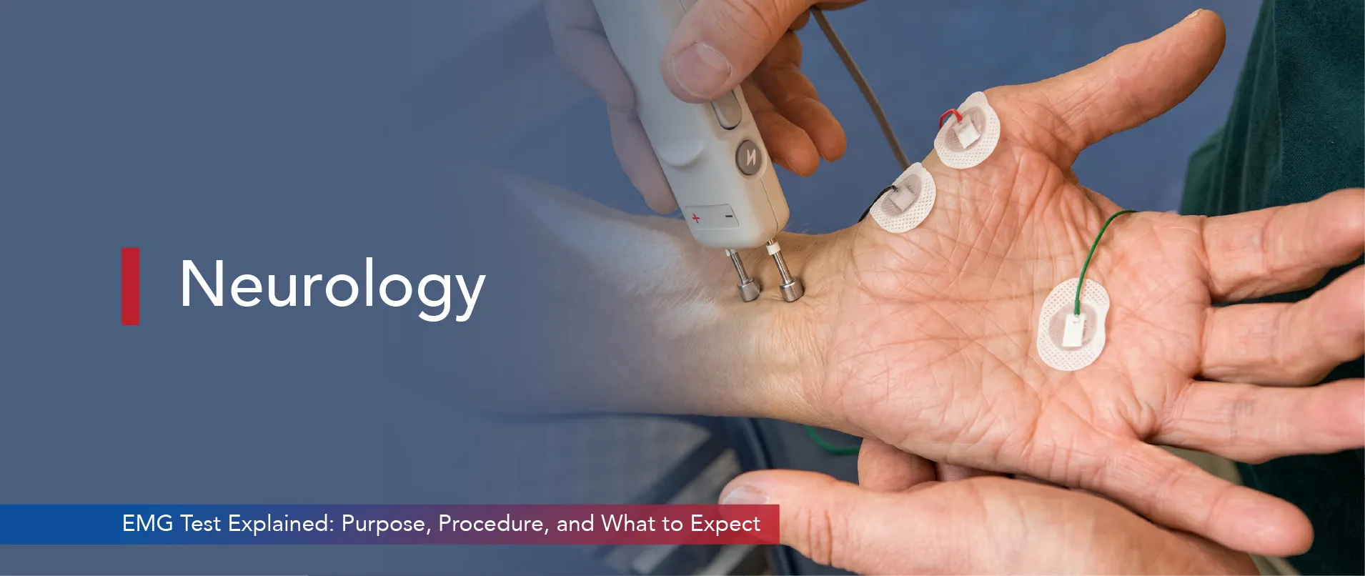
An electromyography (EMG) test is used to analyse the health of your muscles and the nerves that control their movement. These nerves, known as motor neurons, send electrical signals that trigger muscle movement. During the test, these signals are recorded and analysed to help identify problems affecting the muscles or peripheral nerves.
An EMG test records the electrical signals produced by your muscles while they are relaxed and when they constrict. It is combined with a Nerve Conduction Study (NCS), which evaluates how efficiently electrical impulses travel along the nerves.
Together, these tests help doctors distinguish between disorders affecting the muscles, peripheral nerves, nerve roots, or the connection between nerves and muscles.
EMG and NCS are used to evaluate symptoms such as muscle weakness, numbness, tingling, muscle cramps, and unexplained pain. They also help diagnose conditions that affect the nerves and muscles, allowing treatment to begin before the condition progresses.
If you are feeling muscle weakness, persistent numbness, tingling, or unexplained muscle pain, your doctor may recommend an EMG test to determine the underlying cause and guide the most appropriate treatment.
Learning what electromyography is requires a basic understanding of how nerves and muscles interact. Motor neurons transmit electrical signals to muscle fibres. These signals tell the muscle to contract. A motor unit consists of one motor neuron and the muscle fibres it innervates.
An EMG measures the electrical activity generated by muscles. In a healthy muscle, there is minimal electrical activity at rest, while contraction produces a regular pattern of electrical signals.
When a nerve suffers damage, the muscle it controls cannot receive proper signals. The muscle then displays abnormal electrical patterns. EMG testing for nerves helps doctors determine whether the problem originates in the muscle itself or in the controlling nerve.
Neurologists recommend this test for numbness, tingling, or unexplained weakness. The test provides hard data about nerve and muscle function. It removes the guesswork from the diagnostic process entirely.
This test helps doctors to identify a wide range of neuromuscular conditions. Carpal tunnel syndrome is a very common reason for the test. The condition develops when the median nerve gets compressed in the wrist.
EMG test also diagnoses peripheral neuropathies. These conditions damage peripheral nerves, leading to weakness or numbness.
Diabetes often causes this specific type of nerve damage. Doctors also use the test for amyotrophic lateral sclerosis. This progressive disease directly attacks motor neurons.
Myasthenia gravis, a condition causing severe muscle fatigue, also requires an EMG nerve test for an accurate diagnosis.
Muscular dystrophies and spinal disc herniations are other conditions the test can detect. Doctors also diagnose radiculopathies using these results. Radiculopathy occurs when a spinal nerve root becomes compressed.
Peripheral neuropathy is a common neurological disorder that can result from diabetes, infections, autoimmune diseases, and other medical conditions. An early diagnosis of neuromuscular disorders improves patient management. Identifying the specific condition effectively guides the treatment plan.
Knowing what EMG measures helps patients prepare for further procedures. The test records the electrical activity of muscle fibres. A device called an electromyograph captures this activity. It displays the data as waves on a screen.
At rest, a healthy muscle shows minimal electrical activity. When the patient gently contracts the muscle, the electromyograph records the action potentials. Stronger contractions activate more muscle fibres. The electrical signal increases proportionally.
If the muscle or nerve is damaged, the signal pattern shifts. A damaged nerve may send an abrupt signal, producing abnormal sounds and inconsistent patterns on the monitor. The EMG test also tracks how fast electrical signals travel along a nerve. A slower speed points to nerve damage or demyelination. The amplitude of the signal reveals the strength of the muscle response.
These precise measurements help doctors to differentiate between nerve injuries and primary muscle diseases.
A neurologist performs the electromyography test at an advanced clinic. The procedure involves two main parts.
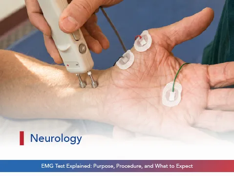
The first part is the nerve conduction study. The doctor places surface electrodes on the skin over the nerve. A small electrical impulse stimulates the nerve. The electrodes then record the signal’s speed and strength. Patients may feel a brief, mild shock during this phase.
The second part of the test is needle electromyography. A thin electrode is inserted into selected muscles to record their electrical activity. You may feel irritation when the needle is inserted and mild discomfort when the muscle is examined.
The patient must relax and then contract the muscle gently. The computer turns the electrical activity into waves on a screen. The doctor reviews these waves in real time. The whole process usually takes about 60-90 minutes.
The electrodes can be affected by body lotions and creams; therefore, avoid them and wear loose clothing so the technician can reach the test sites easily. Once the test is done, you are free to go about your day as normal. An experienced Neurologist in Bangalore ensures the procedure runs smoothly and safely.
Reading electromyography results takes specialised medical knowledge. Normal results show no electrical activity in a resting muscle. Action potentials appear smooth and consistent during contraction. Nerve conduction velocities fall within standard ranges.
Abnormal EMG findings can indicate problems affecting the muscles or nerves. Electrical activity in a resting muscle may suggest an underlying disorder. Changes in the pattern of muscle signals help doctors distinguish between muscle diseases (myopathies) and nerve disorders (neuropathies).
NCS results also determine that slower conduction velocities confirm peripheral nerve damage. A reduced signal amplitude suggests axonal loss. Neurologists combine these results with the physical examination and the patient’s symptoms to determine whether the condition is acute or chronic.
This information is vital for planning effective and timely treatments. Only a trusted neurology hospital in Bangalore with advanced diagnostic equipment can provide highly accurate readings.
An EMG test is a vital diagnostic tool. It reveals the exact cause of muscle weakness and numbness. An accurate diagnosis can prevent further nerve damage and guide proper treatment. Patients should never ignore persistent neurological symptoms.
Sparsh Hospital in Bangalore offers advanced neurological evaluations and progressive diagnostic tests. The hospital provides evidence-based care for all neuromuscular conditions. If you experience unexplained muscle weakness, do not wait. Book an appointment with a specialist at Sparsh Hospital today.
It is a diagnostic procedure that checks the health of muscles and motor neurons. It detects abnormal electrical activity in the muscles.
It measures the electrical activity of muscles at rest and during contraction. It also measures the speed of nerve signals.
The nerve conduction part causes mild tingling. The needle part may cause brief discomfort. However, most patients tolerate the EMG nerve test well.
It diagnoses conditions, such as carpal tunnel syndrome, peripheral neuropathy, muscular dystrophy, and amyotrophic lateral sclerosis.
Consult a Neurologist in Bangalore if you experience persistent muscle weakness, numbness, or tingling. Early evaluation prevents serious complications.
Categories: Neurology



EMG Test Explained: Purpose, Procedure, and What to Expect is available for appointments. Please fill the below form to book an appointment.
Unlock the door to exceptional healthcare, book an appointment with SPARSH Hospital and let your journey to wellness begin.
Copyright © 2026 SPARSH Hospital, A Unit of Shiva & Shiva Orthopaedic Hospital Pvt Ltd.






 Live Chat